


News & Events

New colleague @ BIC!
With the addition of Charlotte Cresens to the light microscopy team we are up to full speed again. Her strong background in super-resolution microscopy (Susana Rocha lab, KUL) will be a great help. Curious? Don't hesitate to pass by and meet her @ the BIC office.
Piezo on AX
The last item of our Nikon AX finally arrived. The stage is now equipped with a 600µm (!) piezo, ideal for fast autofocus operations and z-stacks. Please treat it with care, as all piezo stages it cannot handle high pressure.
Winners BIC Image Contest 2022
Below you can appreciate the winning images of our yearly image contest. Each of these will be used as the desktop wallpaper on the microscope it was acquired on and will stay there till the end of 2022.

Mouse hippocampus
- Cxcl14.Cre x Ai9 P0 hippocampus
- Nuclei in blue (Hoechst), NeuN in magenta, TdTomato in green
- System: Zeiss LSM880 Airyscan

Social Distancing
- System: Zeiss LSM900 Airyscan 2

Neuronal Rosette
- Neuronal Rosettes differentiated from pluripotent stem cells in vitro
- Nuclei in blue (DAPI), NESTIN in green, FOXG1 in magenta, NeuN in light blue
- System: Nikon TiE A1R

Mouse Tibia
- 6 week old Mouse Tibia
- Nuclei in blue (DAPI), stromal cells in orange (LepR), blood vessels in red (Emcn)
- System: Nikon Ti2 AX

Geometric Neurons
- Primary Motor Neurons for ALS research, organized in a geometric pattern
- DIV13
- Nuclei in blue (DAPI), Axons in orange (neurofilament H, AF546), FUS (fused in sarcoma) in purple (AF647)
- System: Nikon TiE A1R

Mitochondria Traffic
- Mitochondria Trafficking in iPSCerived Motor Neurons
- Mitotracker green
- System: Zeiss Cell Discoverer 7

Neurons in Culture
- SIM image
- System: Zeiss Elyra S.1

Cortical Neuron in 3D
- Cortical Neurons projecting to the superior colliculus
- Labeled by retrograde tracing
- 3D-SIM image
- System: Nikon Ti2 nSIM-S

Screening Cells
- System: Operetta CLS

Tracking Memories in the Drosophila Brain
- System: Nikon NiE A1R

Brain Organoids
- Nuclei in purple (DAPI), PAX6 in cyan, SOX2 in orange , GSX2 in green
- System: Nikon Märzhauser Slide Express 2

Cell Reflection
- Reflection of a non-labeled MEF cell
- System: Leica TCS SP5

Motor Neuron in Live Zebrafish
- Live, in vivo, single zebrafish motor neuron
- Zebrafish mutant line homozygous for kif5aa delta exon 27 (mimic of ALS KIF5A mutation obtained via CRISPR gene editing) that carries a motor neuron-specific GAL4 driver
- Expressing UAS construct to co-label the membrane (red), and the mitochondria (green)
- System: Nikon NiE spinning-disk
User satisfaction questionnaire
Your feedback on our services is not only highly appreciated, it's also necessary to keep on improving the functioning of our imaging facility. We would be happy to receive your suggestions, comments and feedback via the link below.
January 2022
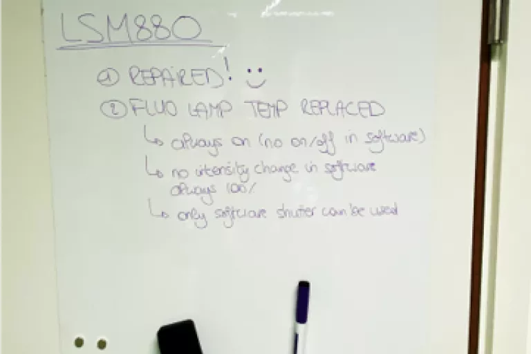
Whiteboards to improve communication
Whiteboards were glued to the door of all microscopy and facilitary rooms in order to facilitate communication about small issues, questions, comments between the BIC and our users, but also between users. Don't hesitate to use them!

Badge accessible cabinet for small parts
Our badge accessible cabinet contains next to a variety of small parts and equipment that can be booked via connect, also an emergency stock of lens cleaning tissue. The cabinet was not only created to provide a safe place for our accessories, it also guarantees 24/7 access for our users. On top of that we can track everything that's inside so nothing get's lost. All credits to Frank Vernaillen (BITS) & Luc Grammet (Mechanisch atelier KUL).

QR code to device manual & scheduler
Each of our high end systems now has a sticker on it with 2 QR codes that guide you directly to either the scheduler page for checking it's availability or to the device manual. Like this you don't have to struggle with printing and you immediately have all the right information at hand.